
Human Heart Outline Drawing at GetDrawings Free download
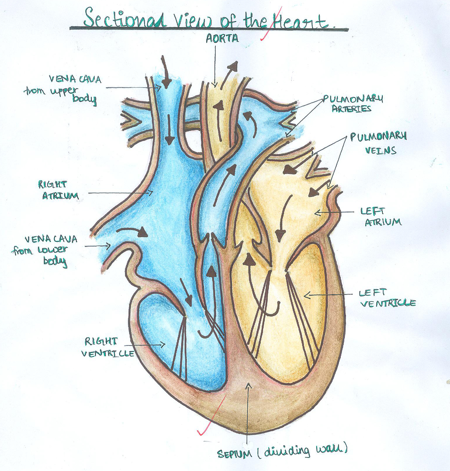
Heart anatomy. The heart has five surfaces: base (posterior), diaphragmatic (inferior), sternocostal (anterior), and left and right pulmonary surfaces. It also has several margins: right, left, superior, and inferior: The right margin is the small section of the right atrium that extends between the superior and inferior vena cava .

Blank Human Heart Diagram learning me Pinterest Human heart
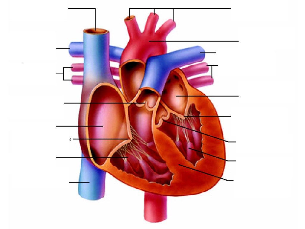
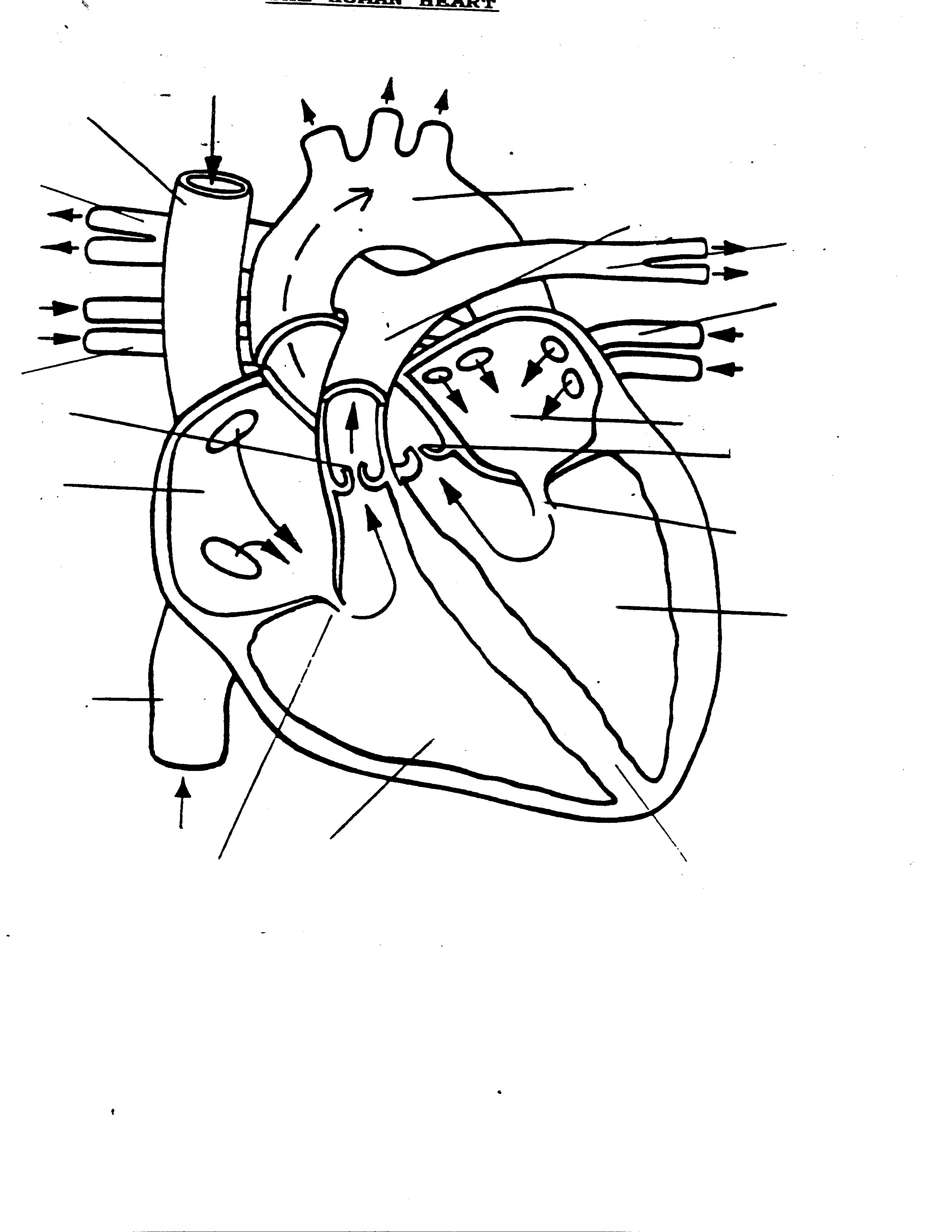
Don't forget to LABEL the parts of the heart on the diagram! 1. Compare the location of the tricuspid and bicuspid. 2. Compare the direction of blood flow in the pulmonary artery to the pulmonary vein. 3. Mitral regurgitation is a heart condition that occurs when the mitral valve does not close fully. Based on your knowledge of the heart.

Simple Human Heart Drawing at GetDrawings Free download
Selecting or hovering over a box will highlight each area in the diagram. For optimal viewing of this interactive, view at your screen's default zoom setting (100%) and with your browser window view maximised. See the Labelling the heart activity for additional support in using this interactive. Parts of the heart
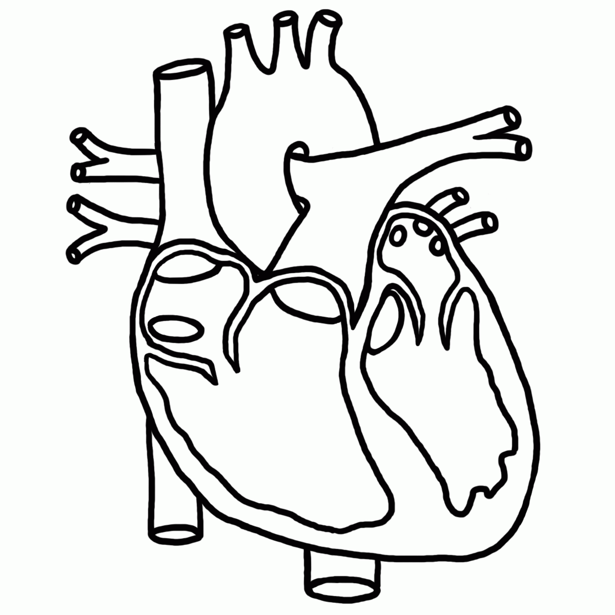
Human Heart Line Drawing at GetDrawings Free download
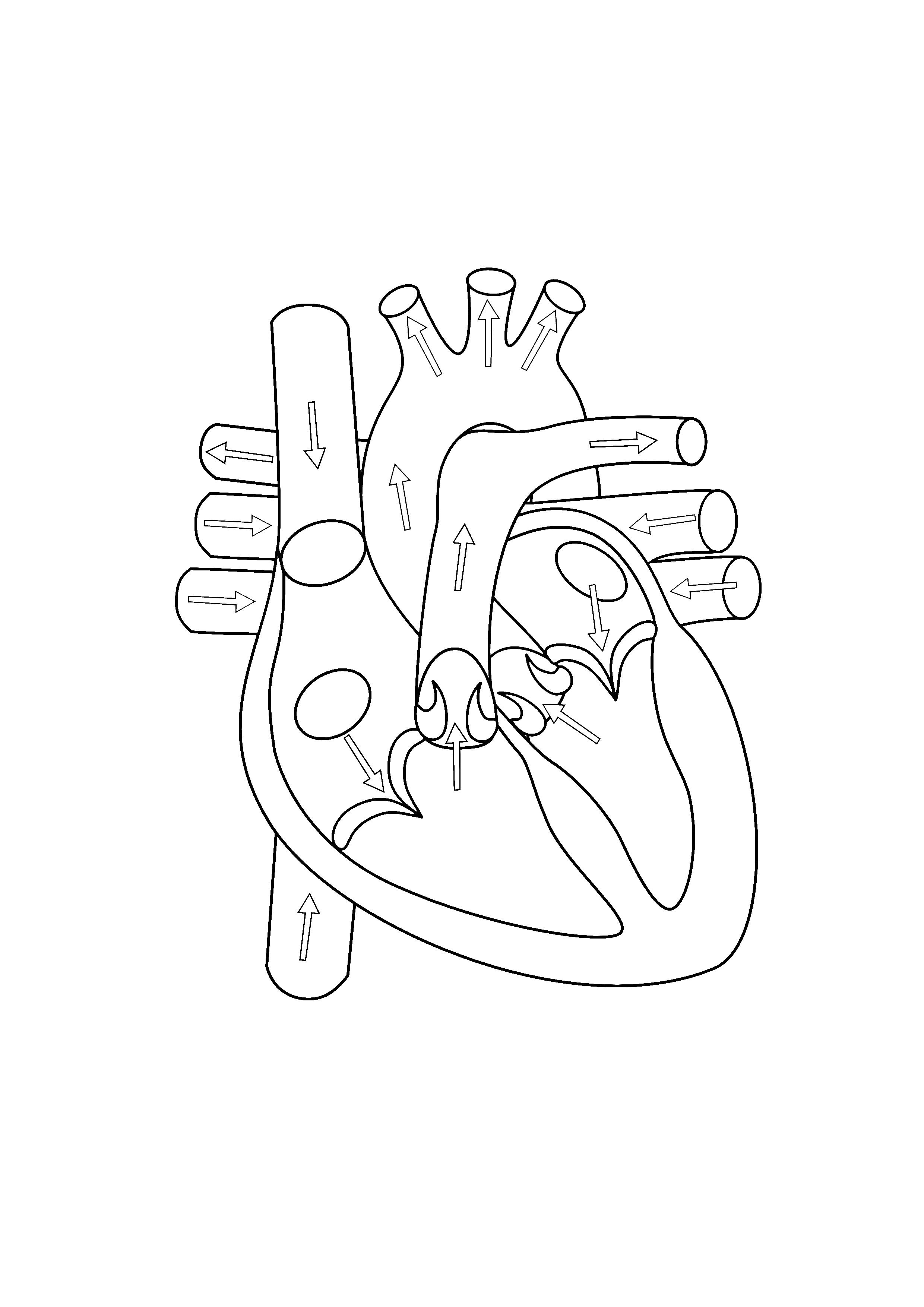
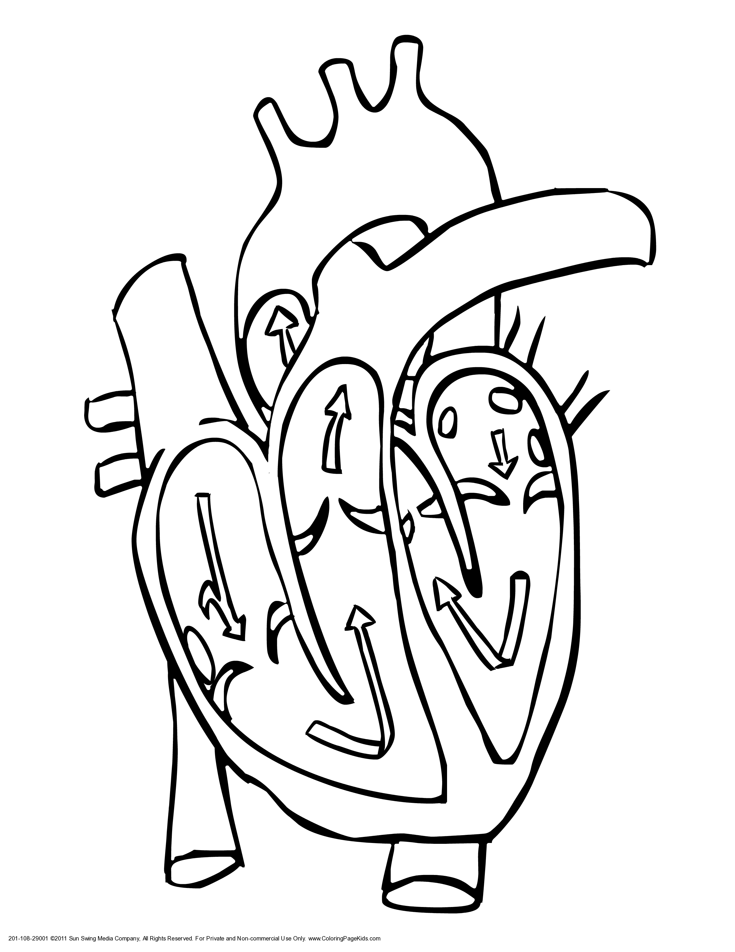
Worksheet showing unlabelled heart diagrams. Using our unlabeled heart diagrams, you can challenge yourself to identify the individual parts of the heart as indicated by the arrows and fill-in-the-blank spaces. This exercise will help you to identify your weak spots, so you'll know which heart structures you need to spend more time studying.

画像をダウンロード heart diagram unlabeled 912873Printable heart diagram unlabeled
This printable blank heart diagram can allow teachers to guide students through the process of identifying and labeling the various parts of the heart. Additionally, an unlabeled heart diagram can serve as a review or assessment tool, helping students reinforce their understanding of the heart's structure and function. Format: PDF. Paper size.
Free Blank Heart Diagram, Download Free Blank Heart Diagram png images
Our blank heart diagram is an excellent visual aid to help students learn about the different parts of the heart, including the atria, ventricles, valves, and major blood vessels.The diagram is also a useful resource for medical professionals who need a quick reference guide to the heart's anatomy. It can be used as a teaching tool or as a.
Blank Printable Heart Diagram Printable World Holiday
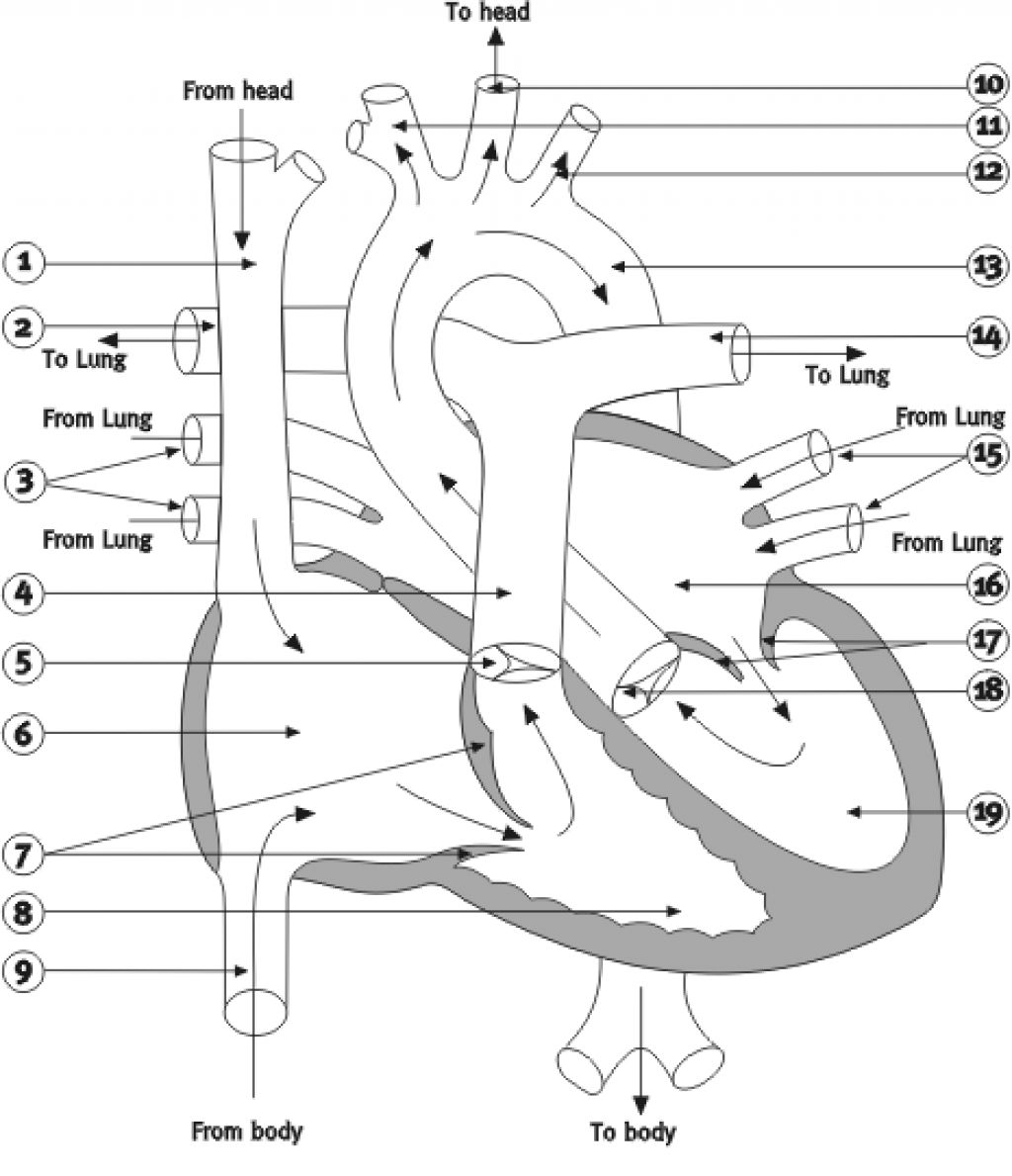
The heart, although a single organ, can be considered as two pumps that propel blood through two different circuits. The right atrium receives venous blood from the head, chest, and arms via the large vein called the superior vena cava and receives blood from the abdomen, pelvic region, and legs via the inferior vena cava.Blood then passes through the tricuspid valve to the right ventricle.
17+ Heart Diagram Templates Sample, Example, Format Download
Diagram of Heart. The human heart is the most crucial organ of the human body. It pumps blood from the heart to different parts of the body and back to the heart. The most common heart attack symptoms or warning signs are chest pain, breathlessness, nausea, sweating etc. The diagram of heart is beneficial for Class 10 and 12 and is frequently.
Blank Heart Diagram Cliparts.co
Heart diagram blank labels are especially valuable for students studying biology, anatomy, or medicine. They are often used in classrooms, laboratories, and online courses to supplement textbooks and lectures. These labeled diagrams provide a hands-on approach to learning, allowing students to actively engage with the material and develop a.
Heart Diagram Unlabeled ClipArt Best
Step 1 and 6 involve a blood vessel, which makes sense as this is how blood enters and exits that side of the heart. Steps 2-5 involve a chamber, valve, chamber, and valve. So if you remember this general pattern, it will help you recall the order in which blood flows through each side of the heart.
Anatomical Heart Coloring Page Coloring Home
Veins return oxygen-poor blood to your heart. Follow these steps to make a chart about the path blood takes through your heart. 1.Cut out the heart diagrams. Glue the heart diagrams in order on the construction paper. 2.Cut out the phrases below. Pair the phrases to make five sentences. 3.Glue each sentence under the matching heart diagram. 1. 2.
Labeled Drawing Of The Heart at GetDrawings Free download
Read the directions on the first page of the "Go With the Flow" printable together. Then have each student complete the activity independently or with a partner. 5. If desired, have students color the diagrams using blue and red crayons or colored pencils to show the oxygen-poor blood and oxygen-rich blood as it travels through the heart.

Printable Blank Unlabeled Heart Diagram glorietalabel
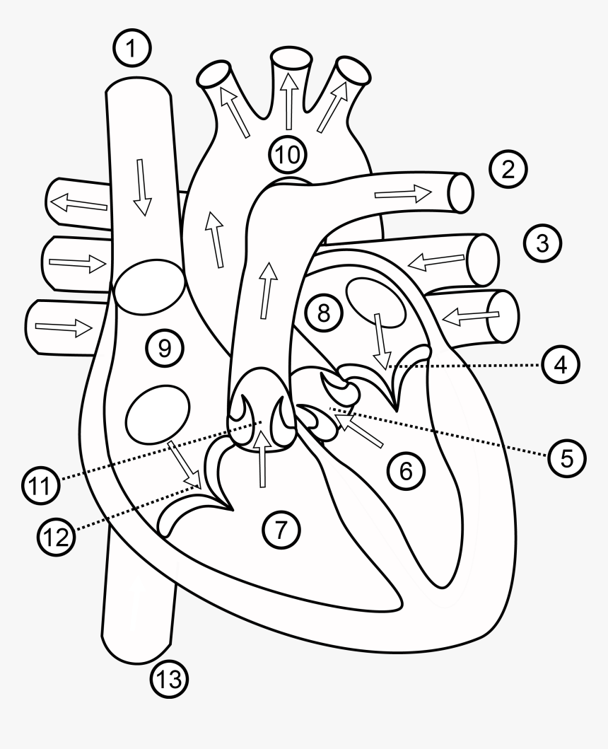
Function and anatomy of the heart made easy using labeled diagrams of cardiac structures and blood flow through the atria, ventricles, valves, aorta, pulmonary arteries veins, superior inferior vena cava, and chambers. Includes an exercise, review worksheet, quiz, and model drawing of an anterior view (frontal section) of the heart in order to.

Human Heart Diagram Unlabeled Tim's Printables
A printable heart diagram is a simple visual aid that highlights the key features of the human heart, such as its chambers, valves, and blood vessels. This type of labeled diagram is ideal for use in classrooms or at home, allowing students to learn about the heart's structure and function in an engaging and interactive way. Format: PDF.
Free Blank Heart Diagram, Download Free Blank Heart Diagram png images
The human heart is located within the thoracic cavity, medially between the lungs in the space known as the mediastinum. Figure 19.2 shows the position of the heart within the thoracic cavity. Within the mediastinum, the heart is separated from the other mediastinal structures by a tough membrane known as the pericardium, or pericardial sac.
Human Heart Drawing Outline At Getdrawings Structure Of Heart Class 7
The heart has three layers. They are the: Epicardium: This thin membrane is the outer-most layer of the heart. Myocardium: This thick layer is the muscle that contracts to pump and propel blood.